Pal histology epithelial tissue lab practical question 2 delves into the fascinating world of epithelial tissue, providing a comprehensive exploration of its structure, functions, and clinical significance. This in-depth analysis equips students with a profound understanding of this essential tissue type, fostering a deeper appreciation for its role in human health and disease.
Epithelial tissue, the outermost layer of organs and cavities, serves as a protective barrier and plays a crucial role in various physiological processes. This practical question guides students through the intricacies of epithelial tissue, empowering them to identify and differentiate between its diverse cell types, unlocking the secrets of its intricate organization and function.
Introduction

Epithelial tissue is one of the four primary tissue types in the human body. It lines the surfaces of the body, including the skin, the lining of the digestive tract, and the lining of the respiratory tract. Epithelial tissue also forms the glands of the body, such as the salivary glands and the pancreas.
Studying epithelial tissue in the histology laboratory is important for several reasons. First, it allows us to learn about the normal structure and function of epithelial tissue. This knowledge is essential for understanding how the body works and how diseases can affect it.
Second, studying epithelial tissue in the histology laboratory can help us to diagnose diseases. By examining the appearance of epithelial cells under a microscope, pathologists can often identify the presence of disease. For example, the presence of abnormal cells in the epithelium of the cervix can be a sign of cervical cancer.
Finally, studying epithelial tissue in the histology laboratory can help us to develop new treatments for diseases. By understanding how epithelial cells function and how they are affected by disease, researchers can develop new drugs and therapies that can target these cells and improve patient outcomes.
Methods
To prepare a slide of epithelial tissue for histological examination, the following steps are typically followed:
- The tissue is collected and fixed in a preservative solution, such as formalin.
- The tissue is embedded in a paraffin wax block.
- The paraffin block is sectioned into thin slices using a microtome.
- The sections are stained with hematoxylin and eosin (H&E) or other stains to make the cells and tissues visible under a microscope.
Results
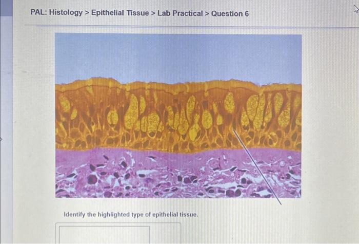
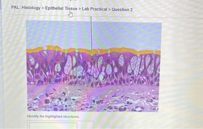
When a slide of epithelial tissue is examined under a microscope, a variety of different cell types can be seen. The most common type of epithelial cell is the squamous cell. Squamous cells are thin, flat cells that are found in the epidermis of the skin and in the lining of the mouth and esophagus.
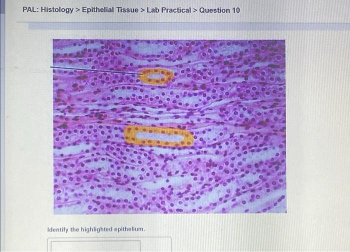
Other types of epithelial cells include cuboidal cells, columnar cells, and transitional cells. Cuboidal cells are cube-shaped cells that are found in the lining of the kidney tubules and the salivary glands. Columnar cells are tall, column-shaped cells that are found in the lining of the stomach and intestines.
Transitional cells are cells that can change shape, and are found in the lining of the bladder.
Discussion

The different types of epithelial tissue have different functions. Squamous cells are adapted to protect the body from the environment. Cuboidal cells are adapted to secrete and absorb substances. Columnar cells are adapted to absorb nutrients and transport materials. Transitional cells are adapted to stretch and contract, which allows them to line the bladder as it fills and empties.
Epithelial tissue is involved in a variety of diseases. For example, squamous cell carcinoma is a type of skin cancer that occurs in the squamous cells of the epidermis. Adenocarcinoma is a type of cancer that occurs in the glandular epithelium.
Transitional cell carcinoma is a type of cancer that occurs in the transitional epithelium of the bladder.
Conclusion

Epithelial tissue is a diverse and important tissue type that plays a vital role in the body. Studying epithelial tissue in the histology laboratory is essential for understanding how the body works, how diseases can affect it, and how to develop new treatments for diseases.
Questions and Answers: Pal Histology Epithelial Tissue Lab Practical Question 2
What is the primary function of epithelial tissue?
Epithelial tissue serves as a protective barrier, lining the surfaces of organs and cavities, and plays a crucial role in various physiological processes, including absorption, secretion, and excretion.
How are epithelial cells classified?
Epithelial cells are classified based on their shape and arrangement, including squamous, cuboidal, and columnar cells, as well as their number of layers, such as simple, stratified, and pseudostratified.
What are the clinical implications of epithelial tissue disorders?
Epithelial tissue disorders can lead to a wide range of health conditions, including skin diseases, respiratory problems, and digestive issues. Understanding the structure and function of epithelial tissue is essential for diagnosing and treating these disorders.